The EEG electrodes(i.e., EOG and ECG) must be placed prior to positioning the participant in the MEG Sensor. This procedure can be carried out during the placement of the MEG head localization coils; prior to digitization.
These are used to identify and monitor eye blinks and saccadic eye movement.
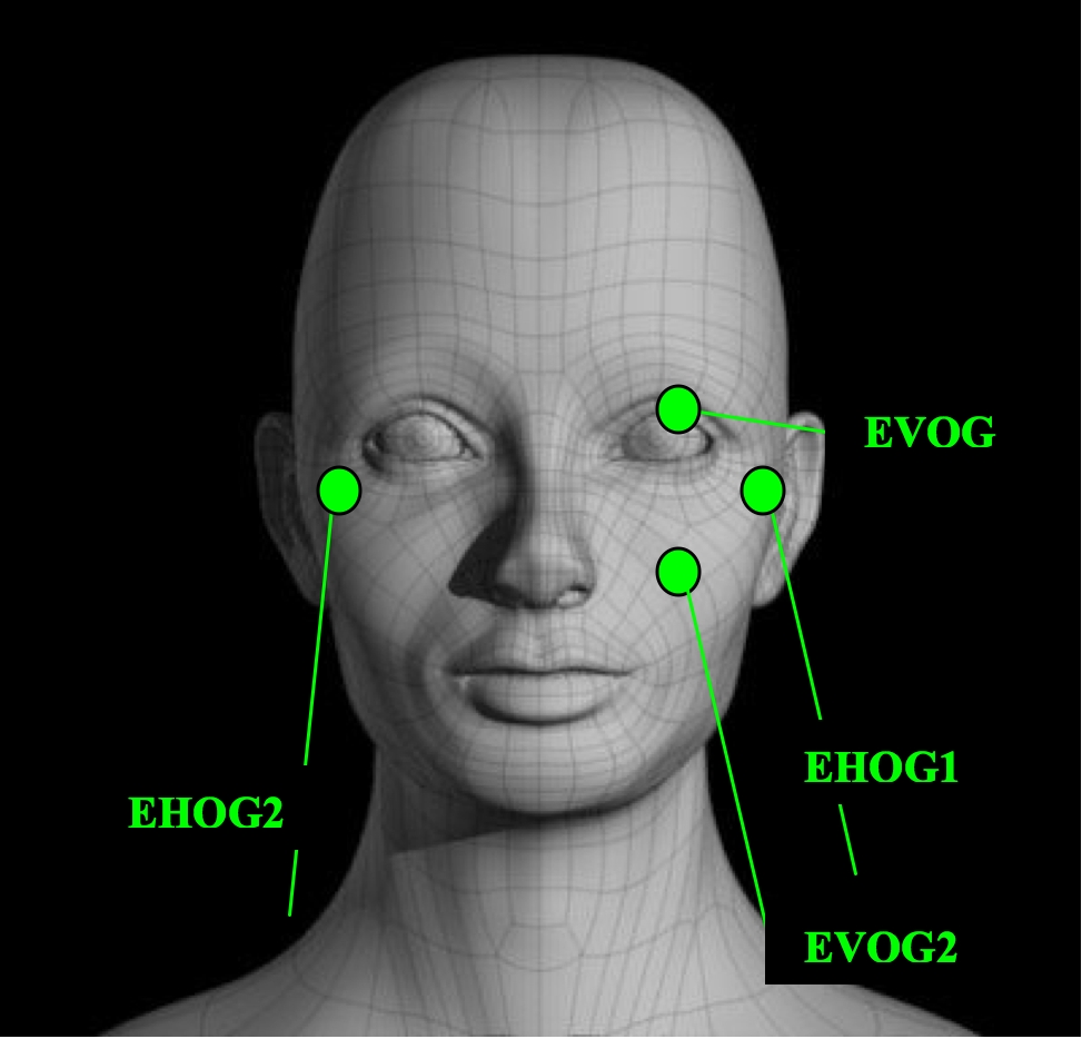
EVOG – Left Eye only. Place electrodes on the orbital ridge centred directly above and below the left eye.
EHOG – Left and Right Eye. Place electrodes at the lateral junction of the upper and lower eyes lid.
Place electrodes as close as possible to the eye without causing discomfort.
- Clean the skin on the cheek near the eyes.
- Attach Large Adhesive Tape (Micropore) to the electrodes.
- Apply Electrolyte Gel through the electrode opening.
- Place the electrodes.
- Press the electrodes onto skin.
- Check the impedances.
- Secure with tape.
This is used to define the electronics common point.
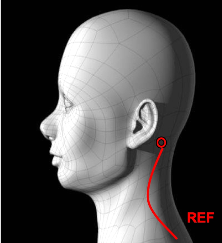
Left Mastoid Reference. , Place the electrode on the left mastoid; which is the bony prominence behind the left ear.
- Clean the skin behind the left ear.
- Attach Adhesive Tape (Micropore) to the superior side of the electrodes.
- Apply Electrolyte Gel through the electrode opening.
- Press the electrodes onto the skin.
- Check the impedances.
- Secure with tape.
These are used to identify and represent the heart’s electrical activity.
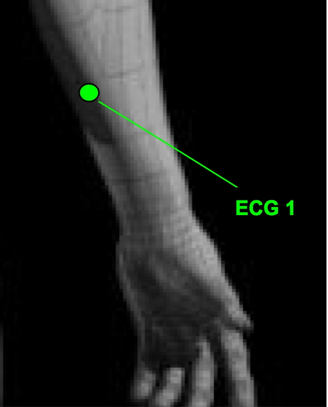
The Left and Right Arm ECG leads are placed on the left and right anterior forearms.
- Clean the skin behind the left and right inner arm.
- Attach Adhesive Tape (Micropore) to the superior side of the electrodes.
- Apply Electrolyte Gel through the electrode opening.
- Place the electrodes onto the skin:
- The negative (ECG1) electrode on the left arm.
- The positive (ECG2) electrode on the right arm.
- Check the impedances.
- Secure with tape.
Electrode | Jack Location |
Mastoids | Ref position (red) |
EHOG - Left / Right Outer Canthus | Bipolar EEG Channel 29 |
EVOG - Supra-Infra Orbital | Bipolar EEG Channel 30 |
ECG - Left/Right Arm | Bipolar EEG Channel 31 |
- EHOG Plug into a bipolar channel.
- EVOG Plug into a bipolar channel.
- Plug the Mastoids into a Linked Electrode Adaptor (Y Connector) then place the single end of the adaptor into the Ref (red) position to link the mastoids together.
- ECG Plug into bipolar channel 31
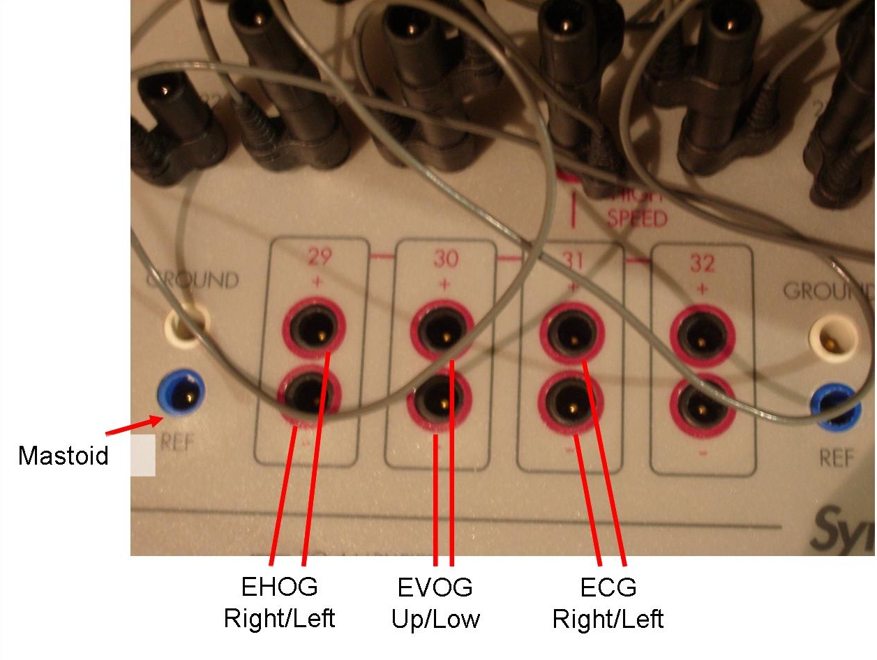
Make sure to plug electrodes in the correct jack box location. Check the montage. Electrode labels on jack box are in the lower right corner of each jack position.